- Home
- Locations & Divisions
- WIVA
- Services
- Interventional Procedures
Request an Appointment
Contact us to discuss any of these procedures or schedule your appointment today!
Recent News
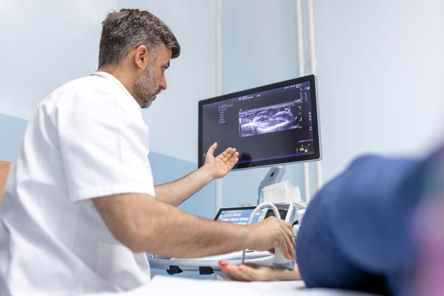
Guided by Imaging
Our patients’ health is our priority, and our trusted Interventional Procedures are designed to provide diagnoses and individualized solutions with minimal disruption to daily life. Casper Medical Imaging & Outpatient Radiology offer a range of Interventional Procedures including interventional oncology, image guided biopsies, and more.
We offer the following minimally invasive procedures of the future, today.
- Biliary Intervention
- Central Line Placement (PICC, etc.)
- Dialysis Intervention
- Stroke Intervention
- Image Guided Biopsy
- Image Guided Gastrostomy Tube Placement
- Inferior Vena Cava Filter Placement and Removal
- Interventional Oncology: Image Guided Thermal Ablation/TACE
- Lower Extremity Venous Disease
- Nephrostomy Tube Placement and Maintenance
- Pain Management (NRB, ESI, Rhizotomy, Facet Injections)
- Pelvic Venous Insufficiency
- Peripheral Arterial Disease (AAA, Balloon Angioplasty / Stent)
- Prostate Artery Embolization
- Uterine Fibroid Embolization
- Varicose Vein Treatment
- Vertebroplasty
- Gonadal Venous Insufficiency
- May Thurner Diagnosis and Treatment.
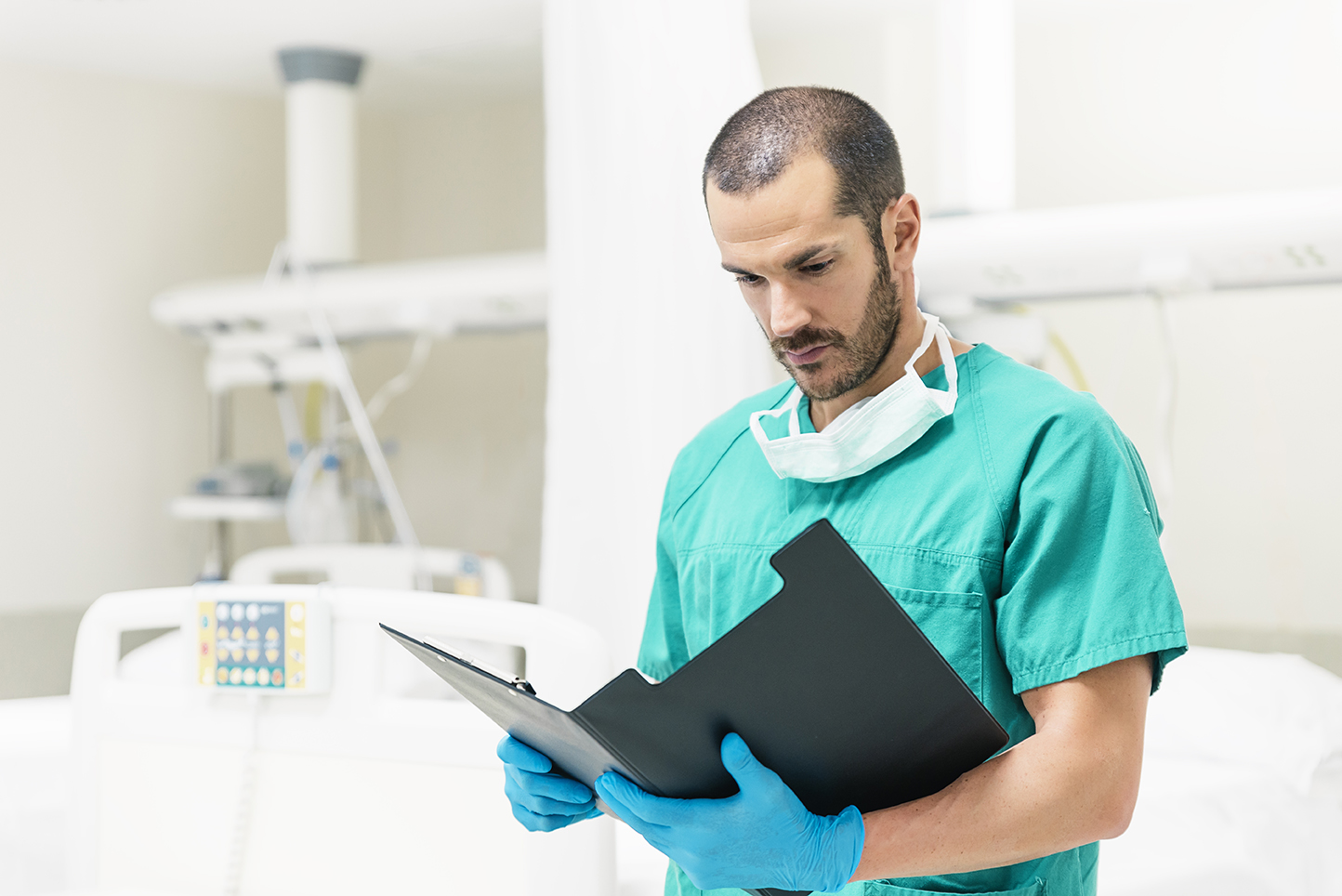

What to Expect
All interventional procedures at WIVA are provided with YOU in mind. Our board-certified and fellowship-trained physicians will ALWAYS meet with you to ensure you get a personal, patient-focused experience, ever time. We’re also proud to have amazing nurses, technologists, and front office staff that are dedicated to making you feel comfortable, heard, and supported throughout your care.
Once your appointment is scheduled, you’ll receive procedure specific instructions to ensure you feel prepared for your procedure.
Testimonials
Exceptional
[The] entire team were exceptional. Some of his team members I knew personally from growing up in Casper which made my experience that much nicer. I felt so good and confident in my care with them.
Saved My Life
[They] took my concerns seriously and treated me immediately. [And] really listened. When people say a doctor saved my life – I truly mean it [..]. I was nearly bed ridden from the pain and swelling and now I am 100% better. My pain is gone and I am even hoping to start riding horses again.