Request an Appointment
Our team is here to serve you every step of the way from baseline screening through problem solving imaging and tissue acquisition. If you have questions, concerns, or would like to speak to a provider please do not hesitate to call and/or schedule an appointment today.
Breast Imaging Excellence
One in eight women, or 12%, will be diagnosed with breast cancer in her lifetime. Despite this fact, we can feel empowered by the opportunity that annual screening provides to detect breast cancer as early as possible. The evidence is clear that early detection of breast cancer improves the odds of survival.
We are proud to be a Designated Comprehensive Breast Imaging Center. This voluntary designation is awarded to centers that demonstrate excellence in breast imaging by earning accreditation in all of the ACR’s breast-imaging modalities.
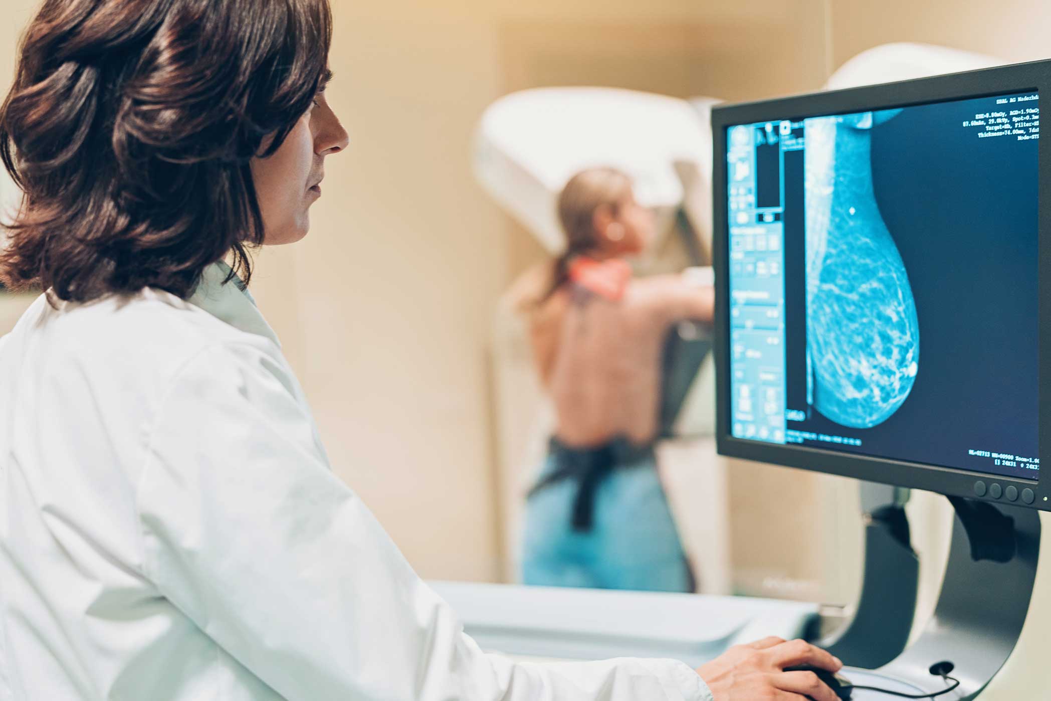
At Casper Medical Imaging & Outpatient Radiology, our focus is you. We prioritize your comfort and care while providing mammography, breast biopsy, and breast ultrasound services so you know where you stand with your health.

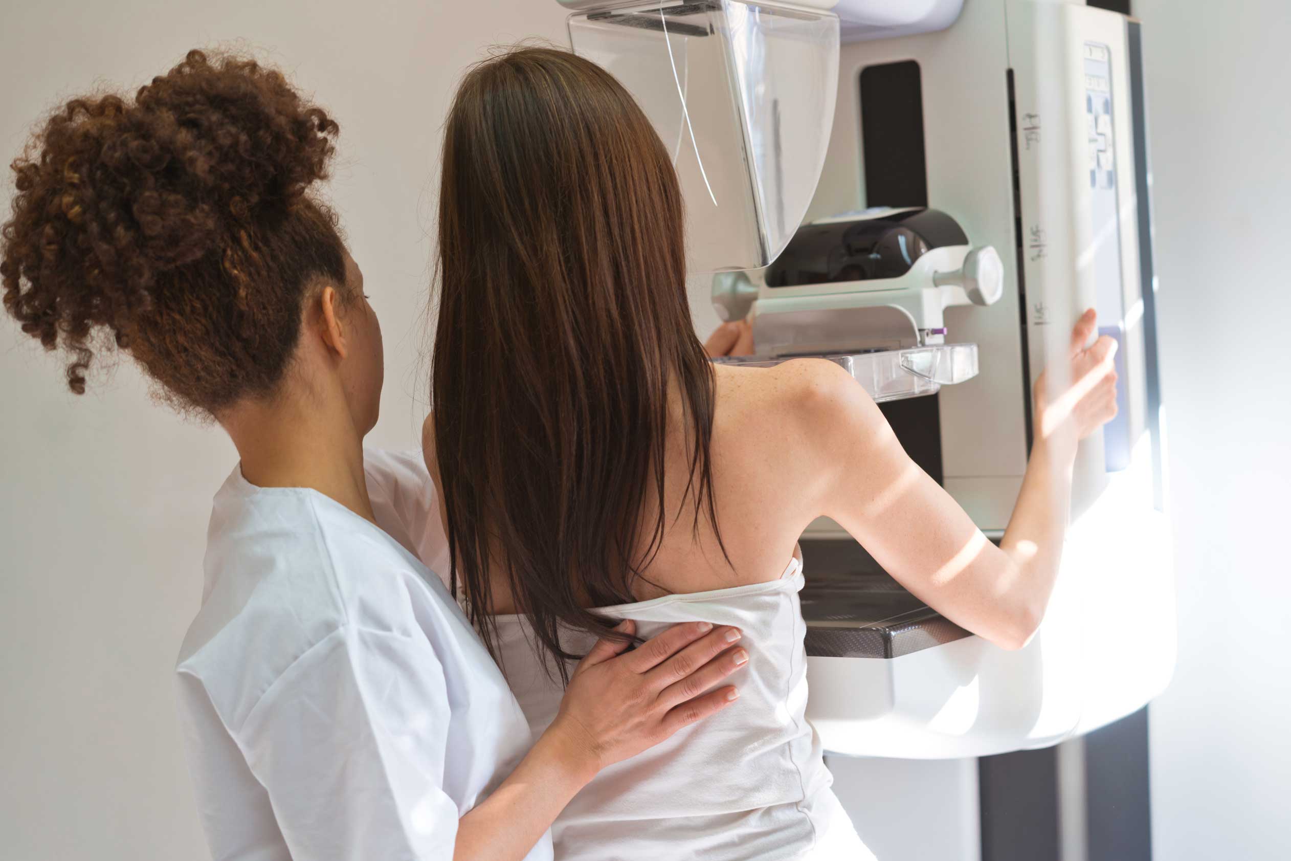
Using a small, safe dose of radiation, mammograms provide the best, most comprehensive images of your breast tissue. We offer standard 2-D view mammography in addition to 3D (or tomosynthesis) to every patient because we know the better your image, the more knowledgeable you’ll be about your breast health.
When a biopsy is recommended, there are many ways to obtain enough tissue to help the pathologist make an accurate diagnosis. Breast lesions can be targeted with specialized mammographic views (termed stereotactic breast biopsy), via ultrasound, or by MRI.
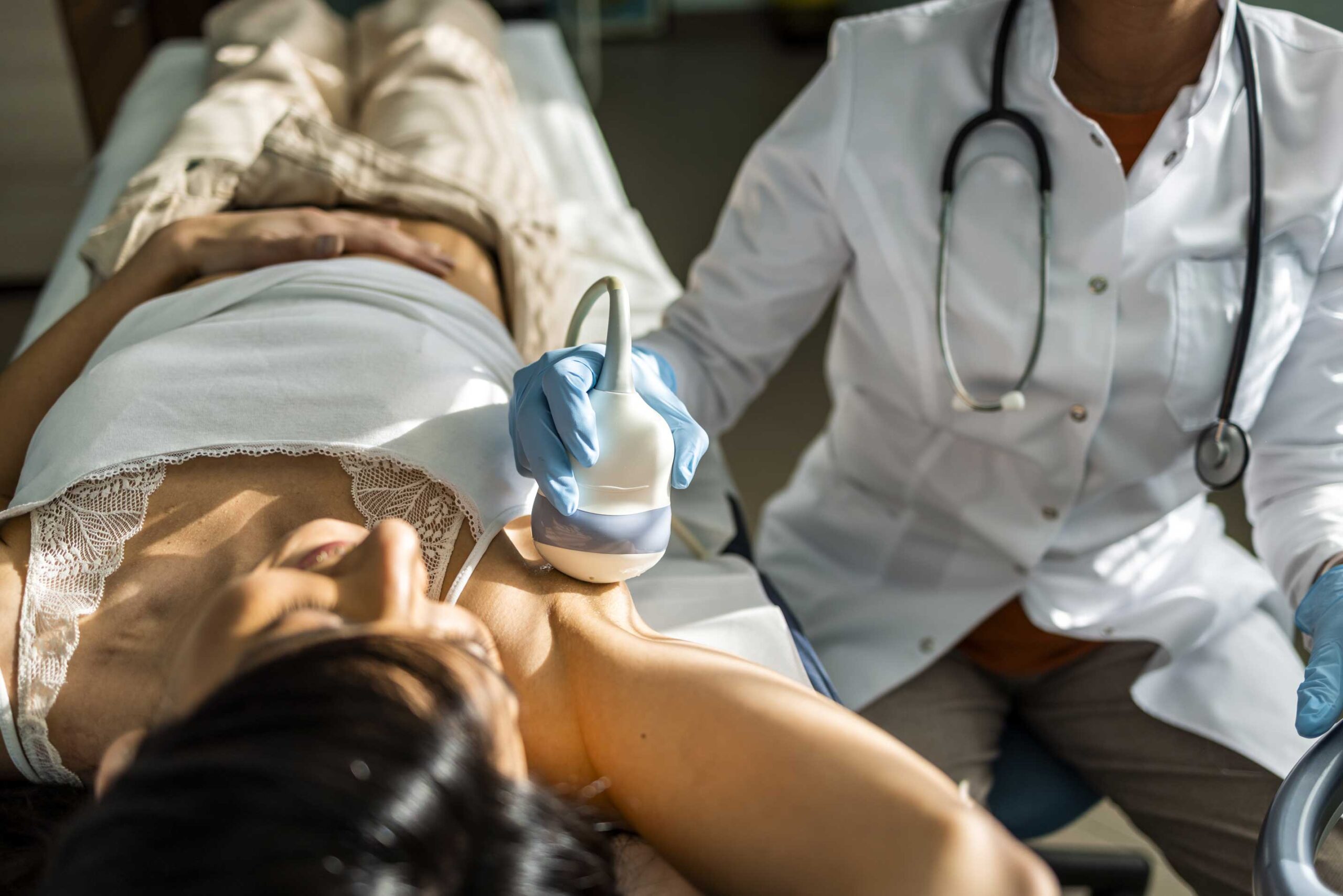
Unlike a mammogram, ultrasound uses sound waves to view the inside of your breast tissue. Ultrasounds are used in conjunction with a mammogram when additional images are needed to help diagnose breast abnormalities.
What to Expect
Your comfort is our number one priority. Throughout the entire process, we aim to make your experience as comfortable as possible, which is why we want you to know what you can expect when you choose CMI & OPR.
Please arrive at your appointment without wearing any deodorant, lotions or powders as they may show on your films and interfere with the quality of your exam.
Upon arrival, your technologist will walk you through your exam. Adequate compression is imperative for a high-quality exam but should be tolerable and your technologist will work with you to ensure you are as comfortable as can be expected. Once your exam is complete, your provider will be sent your results and you will also receive a letter with your results roughly one week from the exam date.
When an abnormality is found, our fellowship trained and board certified radiologists will ensure that you are scheduled for the next stages of diagnosis. Imaging tests that may be required include specialized mammographic views of the breast also known as a “diagnostic mammogram”, a breast ultrasound, a breast MRI, and potentially a biopsy.
FAQ
We don’t like to waste your time. You can expect to spend 20 minutes in your exam – from changing to leaving the facility.
Your provider gets results typically within 24 hours. You will also receive a letter in mail. Our portal also provides access to your imaging results 72 hours after your exam.
This does not mean there is something “wrong.” It means there has been an area of interest identified on your mammogram which requires additional images, or a Diagnostic Mammogram. If that’s the case, we’ll get you scheduled for additional images. You can expect that visit to take 30-45 minutes so the radiologist can review your imaging at the time of your exam. The radiologist will then determine if there is a need for further imaging such as an ultrasound, which will take place during the same visit. Our onsite radiologists are available to you during this exam should you have questions or concerns about your imaging and often times we can provide you with real time recommendations and results.
Other times, the radiologist may recommend a short term follow up, usually within in six months. This category is called BIRADS 3 and may require imaging over a two-year period to ensure the area is stable.
When it comes to breast biopsies there are many ways to obtain enough tissue to help the pathologist make an accurate diagnosis. Breast lesions can be targeted with a specialized mammography unit, termed stereotactic breast biopsy. Other methods include ultrasound, or by MRI.
1) Stereotactic breast biopsies are performed in compression, like a mammogram. Once the breast is in compression, images are acquired and the lesion is localized. The skin overlying the lesion is then cleansed, the skin and deeper tissues are numbed with local anesthesia, and a small incision (roughly the size of a pea) is made in the skin to allow the needle to pass. Once the needle is in place and the sample is obtained, a very small marker, called a clip, is left in the biopsy cavity to identify the biopsy location should more tissue need to be removed at a later date. Complications are rare and you can get back to your normal life immediately.
2) Ultrasound guided breast biopsies are performed without compression and use a small probe to locate the lesion. Once the lesion is localized, the skin overlying the lesion is cleansed, the skin and deeper tissues are numbed with local anesthesia, and a small incision (roughly the size of a pea) is made in the skin to allow the needle to pass. Once the needle is in place and the sample is obtained, a very small marker, called a clip, is left in the biopsy cavity to identify the biopsy location should more tissue need to be removed at a later date. Again, complications are rare, and you can get back to your normal life immediately.
3) MRI guided breast biopsies are performed with a mild degree of compression, and the breast is placed into a special depression in the MRI table. Breast imaging and biopsies are performed in the prone position, laying on your front side and face down. A small plastic grid is placed on the side of the involved breast and images are taken without and with IV contrast. Once the lesion is localized, the skin overlying the lesion is cleansed, the skin and deeper tissues are numbed with local anesthesia, and a small incision (roughly the size of a pea) is made in the skin to allow the needle to pass. Once the needle is in place and the sample is obtained, a very small marker, called a clip, is left in the biopsy cavity to identify the biopsy location should more tissue need to be removed at a later date. Again, complications are rare, and you can get back to your normal life immediately.
Very Little Discomfort
The technologist was gentle and considerate and there was very little discomfort in the process of the mammogram. The results were immediately reviewed with me.