Request an Appointment
If you have questions, concerns, or would like to speak to a provider please do not hesitate to call and/or schedule an appointment today.
Recent News
Your image is Our Focus
-
X-Ray
-
An x-ray (radiograph) is a noninvasive medical test that helps physicians diagnose and treat medical conditions. Imaging with x-rays involves exposing a part of the body to a small dose of ionizing radiation to produce pictures of the inside of the body. X-rays are the oldest and most frequently used form of medical imaging. A bone x-ray makes images of any bone in the body, including the hand, wrist, arm, elbow, shoulder, spine, pelvis, hip, thigh, knee, leg (shin), ankle or foot.
Chest x-ray uses a very small dose of ionizing radiation to produce pictures of the inside of the chest. It is used to evaluate the lungs, heart and chest wall and may be used to help diagnose shortness of breath, persistent cough, fever, chest pain or injury. It also may be used to help diagnose and monitor treatment for a variety of lung conditions such as pneumonia, emphysema and cancer. Because chest x-ray is fast and easy, it is particularly useful in emergency diagnosis and treatment.
Additional Information
What to Expect -
Ultrasound
-
Ultrasound imaging, also called sonography, involves exposing part of the body to high-frequency sound waves to produce pictures of the inside of the body. Ultrasound exams do not use ionizing radiation (as used in x-rays). Because ultrasound images are captured in real-time, they can show the structure and movement of the body’s internal organs, as well as blood flowing through blood vessels.
Conventional ultrasound displays the images in thin, flat sections of the body. Ultrasound examinations can help to diagnose and treat a variety of medical conditions and to assess organ damage following illness. Ultrasound is used to help physicians evaluate symptoms such as pain, swelling, and infection and is a useful way of examining many of the body’s internal organs.
Additional Information
What to Expect -
CT Scan
-
Computed tomography (CT) is a diagnostic imaging test used to create detailed images of internal organs, bones, soft tissue and blood vessels. Like an X-ray, it shows structures inside your body but in greater detail. For example, body structures overlap on regular X-rays and cause many things to remain hidden from view. A CT shows the details of each of your organs for a clearer and more precise view. In short, A CT is fast, minimally invasive, and accurate!
Another term for CT scan is CAT scan. CT stands for “computed tomography,” while CAT stands for “computed axial tomography.” But these two terms describe the same imaging test.
Additional Information
What to Expect
FAQ -
DEXA
-
Bone densitometry, also called dual-energy x-ray absorptiometry, DEXA, uses a very small dose of ionizing radiation to produce pictures of the inside of the body (usually the lower, or lumbar, spine and hips) to measure bone loss. It is commonly used to diagnose osteoporosis, to assess an individual’s risk for developing osteoporotic fractures. DEXA is simple, quick and noninvasive.
Additional Information
What to Expect
FAQ -
LOW DOSE LUNG CANCER SCREENING
-
The only proven method of screening for lung cancer is by low dose CT. All it takes is 10 minutes of your time, no needles, and no medications. Patients with the following risk factors should be screened annually for lung cancer: We offer low dose lung cancer screening to every patient at risk for lung cancer. A few ways to know if you are at risk for lung cancer include a 20 pack-year history of smoking, you still smoke or quit within the past 15 years, and you’re between 55 and 80 years old. If that’s you, you schedule your annual low dose lung CT scan to check for cancer!
-
FLUOROSCOPY
-
Fluoroscopy is an imaging modality that uses x-rays to allow real-time visualization of body structures. During fluoroscopy, x-ray beams are continually emitted and captured on a screen, producing a real-time, dynamic image. This allows for dynamic assessment of anatomy and function. With the ability to assess these functions in real time, you may be given contrast. Contrast materials, also known as contrast agents or contrast media, are used to temporarily change the way x-rays or other imaging tools interact with the body. Contrast materials are not dyes that permanently discolor internal organs. They are substances that are used to accentuate specific structures when looking at a body image. Contrast can be an iodine-based material, barium-sulfate, gadolinium, or saline and air mixture that can be swallowed or injected intravenously. Contrast helps to distinguish, or “contrast,” between organs, tissues, bones, or blood vessels during your imaging exam3. Studies performed under fluoroscopy can include esophagrams, upper and lower GI studies as well as many types of injections where the real time imaging helps guide needle placement.
An x-ray (radiograph) is a noninvasive medical test that helps physicians diagnose and treat medical conditions. Imaging with x-rays involves exposing a part of the body to a small dose of ionizing radiation to produce pictures of the inside of the body. X-rays are the oldest and most frequently used form of medical imaging. A bone x-ray makes images of any bone in the body, including the hand, wrist, arm, elbow, shoulder, spine, pelvis, hip, thigh, knee, leg (shin), ankle or foot.
Chest x-ray uses a very small dose of ionizing radiation to produce pictures of the inside of the chest. It is used to evaluate the lungs, heart and chest wall and may be used to help diagnose shortness of breath, persistent cough, fever, chest pain or injury. It also may be used to help diagnose and monitor treatment for a variety of lung conditions such as pneumonia, emphysema and cancer. Because chest x-ray is fast and easy, it is particularly useful in emergency diagnosis and treatment.
Additional Information
What to Expect
Ultrasound imaging, also called sonography, involves exposing part of the body to high-frequency sound waves to produce pictures of the inside of the body. Ultrasound exams do not use ionizing radiation (as used in x-rays). Because ultrasound images are captured in real-time, they can show the structure and movement of the body’s internal organs, as well as blood flowing through blood vessels.
Conventional ultrasound displays the images in thin, flat sections of the body. Ultrasound examinations can help to diagnose and treat a variety of medical conditions and to assess organ damage following illness. Ultrasound is used to help physicians evaluate symptoms such as pain, swelling, and infection and is a useful way of examining many of the body’s internal organs.
Additional Information
What to Expect
Computed tomography (CT) is a diagnostic imaging test used to create detailed images of internal organs, bones, soft tissue and blood vessels. Like an X-ray, it shows structures inside your body but in greater detail. For example, body structures overlap on regular X-rays and cause many things to remain hidden from view. A CT shows the details of each of your organs for a clearer and more precise view. In short, A CT is fast, minimally invasive, and accurate!
Another term for CT scan is CAT scan. CT stands for “computed tomography,” while CAT stands for “computed axial tomography.” But these two terms describe the same imaging test.Additional Information
What to Expect
FAQ
Bone densitometry, also called dual-energy x-ray absorptiometry, DEXA, uses a very small dose of ionizing radiation to produce pictures of the inside of the body (usually the lower, or lumbar, spine and hips) to measure bone loss. It is commonly used to diagnose osteoporosis, to assess an individual’s risk for developing osteoporotic fractures. DEXA is simple, quick and noninvasive.
Additional Information
What to Expect
FAQ
The only proven method of screening for lung cancer is by low dose CT. All it takes is 10 minutes of your time, no needles, and no medications. Patients with the following risk factors should be screened annually for lung cancer: We offer low dose lung cancer screening to every patient at risk for lung cancer. A few ways to know if you are at risk for lung cancer include a 20 pack-year history of smoking, you still smoke or quit within the past 15 years, and you’re between 55 and 80 years old. If that’s you, you schedule your annual low dose lung CT scan to check for cancer!
What to Expect
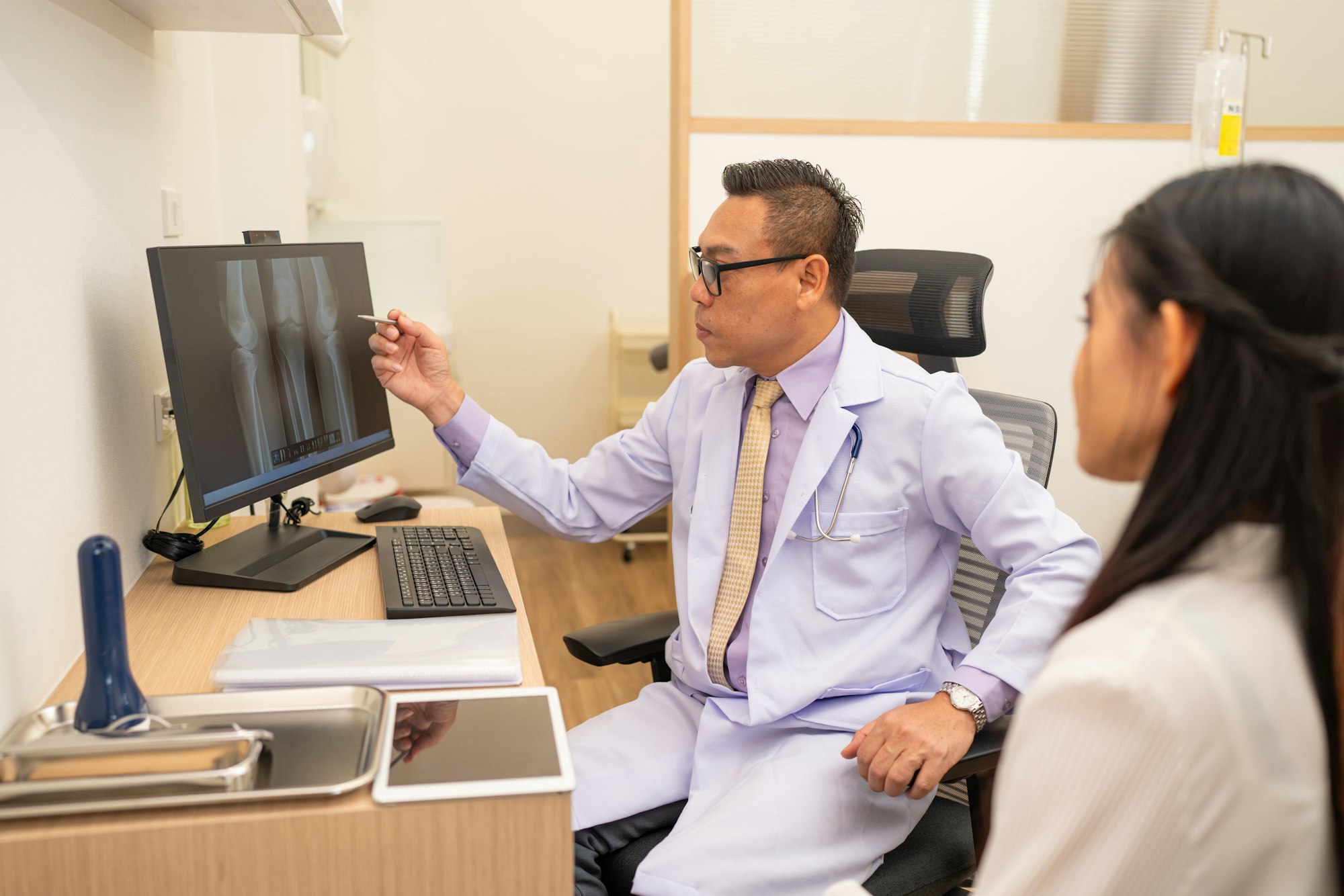
All of our diagnostic imaging services are provided with you, the patient in mind. Regardless of your service, we strive to ensure you are informed and comfortable every step of the way. To that end, here’s briefly what you can expect from your service.
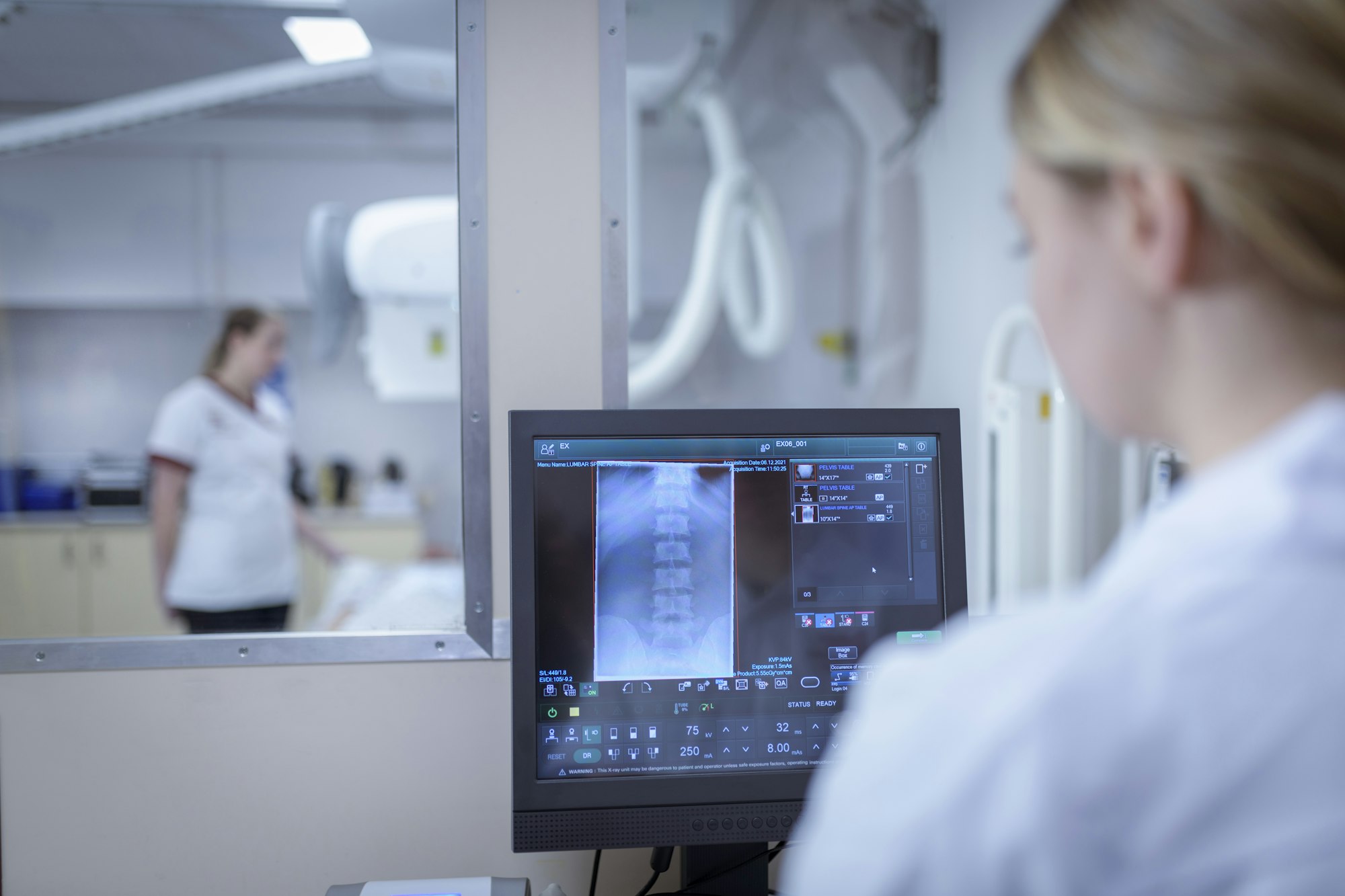
X-ray: Depending on what part of your body will require imaging, you may be asked to change into a gown, as some clothing materials can show up on our high-quality imaging. Some exams can be performed standing and other times you may be asked to lay on the imaging table. The images are acquired digitally, and your technologist will review them immediately upon exposure. Once your exam is complete the images will be reviewed by a board certified, and fellowship trained radiologist and a report will be sent to your provider usually within 24 hours or less.
Ultrasound: This procedure requires bare skin and the application of gel-like solution. While you won’t be required to fully undress, ensure the area being scanned is accessible. Once the solution is applied to the skin, our technologist will use a want to gently glide over the area to view the inside of the body. One the procedure is complete, the technologist will provide a wipe or towel so you can remove the gel solution.
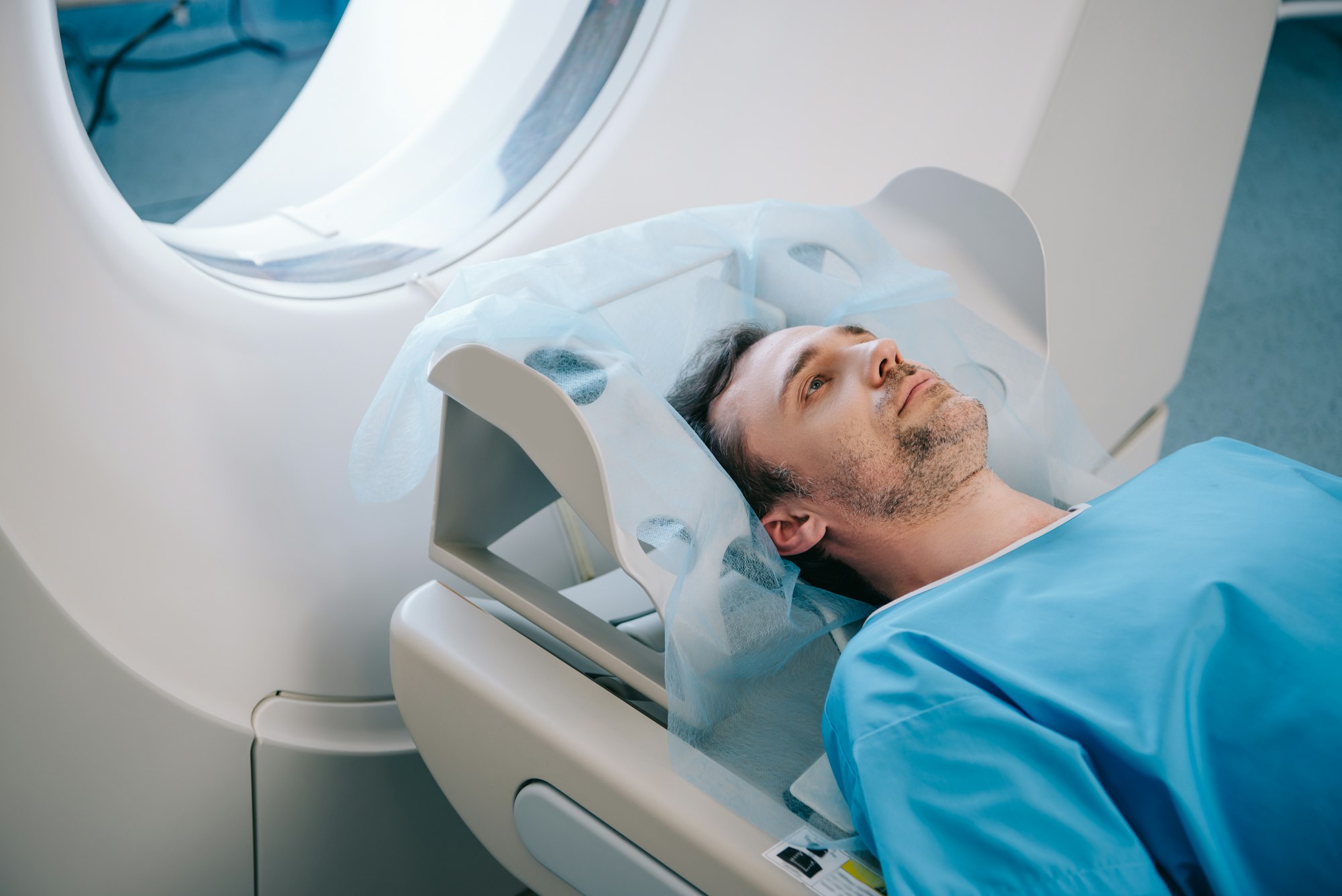
CT – For a CT scan with contrast, you will receive an IV (intravenous line) and injection of contrast (or dye) into your vein to highlight certain areas of your body on the scan. This dye and can make you feel flushed or give you a metallic taste in your mouth. You may also be given an oral contrast to highlight your intestines. Both improve the visibility of specific tissues, organs or blood vessels and help healthcare providers diagnose several medical conditions. IV contrast agents usually flush from your system (when you urinate) within 24 hours.
During a CT, you’ll usually lie on your back on a table (like a bed). When the scan begins, the bed will slowly move into the doughnut-shaped scanner. At this point, you’ll need to stay as still as possible because movement can blur the images.
You may also be asked to hold your breath for a short period of time, usually fewer than 15 to 20 seconds. The scanner takes pictures of the area your healthcare provider needs to see. Unlike an MRI scan, a CT scan is silent. When the exam is over, the table moves back out of the scanner.
DEXA – For your Dexa scan, you will be asked to refrain from taking any oral calcium or multivitamins for one week prior to your imaging. Because the scan is specifically measuring your bone density, calcium tablets can overlay important anatomy and skew results. The most common area to be scanned for bone density is the hips and lumbar spine, or low back. Because metal can also alter results, you will be asked to wear loose, comfortable clothing without any metal around your hips, waist or torso. If you arrive and this is not the case, a gown will be provided. A brief questionnaire pertaining to your bone and personal health will be reviewed and our team will also measure your current height and weight. Once the scan begins and your personal criteria assessed, your hips and spine will be imaged. After images are obtained and analyzed, a report will be generated with specific information regarding normal bone, osteopenia or osteoporosis.
FAQ
CT Scan
CT scans can detect various injuries and diseases such as cancers, benign tumors, blood clots, internal bleeding, bowel disorders (Crohn’s disease, appendicitis, diverticulitis, blockages), kidney stones, brain injuries, broken bones, etc.
Hydration is encouraged leading up to your appointment. Wear comfortable clothes and remove any metal jewelry or clothing with zippers, snaps, metal buttons or grommets (you may be given a hospital gown to wear).
Most CT exams are complete in fewer than 10 to 15 minutes.
Our radiologists provide results to the ordering provider within 24 to 48 hours.
CT scans are generally safe, even for children. If your child requires a CT scan, automatic adjustments are made to lower the ensure a lower dose of radiation exposure. Like X-rays, CT scans use a small amount of ionizing radiation to capture images. Possible risks of radiation include:
Cancer risk: Imaging using radiation, such as X-rays and CT scans, in theory, may cause a slight increase in your risk of developing cancer. The difference is too tiny to measure effectively.
Allergic reactions: Occasionally, people have an allergic reaction to the contrast agent. This could be a minor or serious reaction.
CT scans of your pelvis and abdomen can expose the developing fetus to radiation, but it’s not enough to cause harm. However, unless deemed medically necessary, we’d recommend waiting until after you’ve delivered to get a CT scan of your pelvis or abdomen. CT scans in other parts of your body don’t put the fetus at any risk.
Cancer risk: Imaging using radiation, such as X-rays and CT scans, in theory, may cause a slight increase in your risk of developing cancer. The difference is too tiny to measure effectively.
Allergic reactions: Occasionally, people have an allergic reaction to the contrast agent. This could be a minor or serious reaction.
DEXA
A DEXA scan requires little to no special preparation. Tell your doctor and the technologist if there is a possibility you are pregnant or if you recently had a barium exam or received an injection of contrast material for a CT or radioisotope scan. Leave jewelry at home and wear loose, comfortable clothing. You may be asked to wear a gown. You should not take calcium supplements for at least 24 hours before your exam.
Exemplary
The ultrasound tech was exemplary. She was kind, attentive, and did a stellar job helping me to feel comfortable during what can only be called a very uncomfortable procedure.
Very Friendly & Knowledgeable
CMI was very efficient in all respects. The check in was quick and I maybe waited 5 minutes before being taken back for my X-ray. Very friendly and knowledgeable rad.tech. – Happy with my experience.
Professional & Caring
The staff and technologists were professional and very caring. This was my first CT with contrast and I have [some allergies] so they were very tentative and showed genuine concern and checked on me throughout to make sure I didn’t have a reaction.